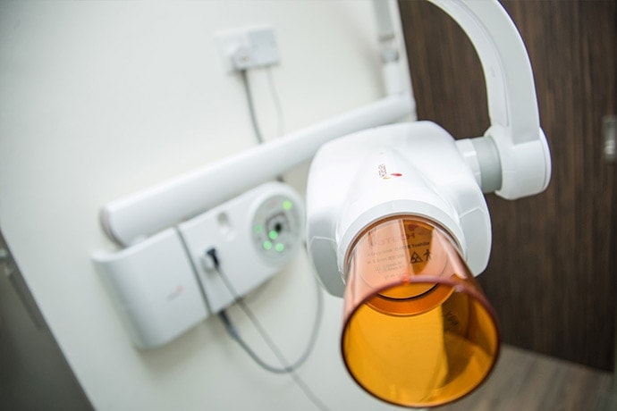
Digital X-Ray Facilities (Cone-Beam Computed Tomography)

In LivingSmile Dental, we use the latest dental digital imaging X-ray technique called Cone-Beam Computed Tomography. Instead of developing X-ray film in a darkroom, the X-rays are sent directly to a computer and can be viewed on screen, stored, or printed out. Our Pax-I 3D Smart CBCT is capable in producing superb 2D panoramic images, as well as 3D imaging and Cephalometric to meet all diagnostic needs.

Our Pax-I 3D Smart CBCT produces FOV 12×9 images can provide the most optimal information for oral diagnosis fully covering both maxillary and mandibular structures, including the 3rd molar region in a single scan. It is suitable for most oral surgery cases, as well as multiple implant surgery.
Benefits of using this new digital imaging technology:
- The technique uses less radiation than the typical X-ray and there is no waiting time for the X-rays to develop — the images are available on screen a few seconds after being taken.
- The image taken, of a tooth for example, can be enhanced and enlarged many times its actual size on the computer screen, making it easier for your dentist to show you where and what the problem is.
- If necessary, images can be electronically sent to another dentist or specialist — for instance, for a second opinion on a dental problem.